RESEARCH
Beginning of Research
Studies on stem cells began at the Butantan Institute (Brazil) in 2004 by researcher Prof. Dr. Irina Kerkis.

Prof. Dr. Irina Kerkis holds:
- Degree in Biology and Chemistry from Tomsk Federal University (1978)
- Master's in Biology and Chemistry from Tomsk Federal University (1981)
- PhD in Cytogenetics from the Institute of Cytology and Genetics (1989)
- Habilitation in Biological Sciences from the Russian Academy of Sciences in 1994
Since 2004 she has been director of the Genetics Laboratory at the Butantan Institute and CNPQ level VI Researcher.
Prof. Irina and her research group have extensive experience in obtaining, characterizing and large-scale production of various types of mesenchymal stem cells. Her current focus is translational medicine using immature dental pulp stem cells in neurodegenerative and hematopoietic diseases.
She has published more than 110 scientific articles, 20 book chapters and produced five patents, three of which have already been granted.
Pre-Clinical Research
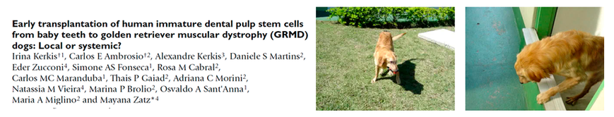
In 2008 the research group of Prof. Dr. Irina Kerkis published important data on muscular dystrophy: Golden retriever muscular dystrophy (GRMD) represents the best available animal model for therapeutic studies aimed at treating Duchenne muscular dystrophy (DMD), a hereditary, lethal and degenerative disease.
CONCLUSION:
This study concluded that systemic (intravenous) administration of mesenchymal stem cells is effective for treating this type of disease, believing that the beneficial effects observed in the animals likely occurred due to the immunomodulatory effects of the cells, in addition to their capacity for regeneration and differentiation. The animal that received monthly intravenous injections remained clinically stable after 25 months of age, which is relatively rare due to the pre-existing pathology, remaining healthy until the time (6 years of age – time of publication), with no signs of tumor formation, according to the researchers' report.
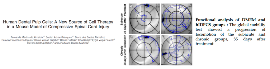
In 2011 the research group of Prof. Dr. Irina Kerkis published important data on Spinal Cord Injury:
In this study, the effects of mesenchymal cells (HDPCs) were evaluated in a mouse model of compressive spinal cord injury (SCI). The cells were transplanted 7 days or 28 days after injury, in order to compare recovery when treatment is applied in subacute or chronic phase.
CONCLUSION:
This study concluded that: Animals that received HDPC transplantation showed better spinal cord preservation than DMEM (control) groups, higher levels of trophic factor expression in tissue, better neuron organization and the presence of many axons myelinated by Schwann cells or oligodendrocytes, in addition to some intact neurons with a healthy appearance. Animals that received the therapy demonstrated an improvement in locomotor functional capacity. Thus, based on these findings, the group proposes that HDPCs are possible candidates for Central Nervous System disorders in Humans.
More Pre-Clinical Data (Multiple Sclerosis Model)
Results of veterinary therapeutic efficacy in canine distemper virus in dogs (= 20), a demyelination model similar to multiple sclerosis etiology: > 90% of patients demonstrate partial recovery after the third transplant.
Allogeneic transplantation of canine CTIPD provides complete and stable recovery in neurological and motor function in dogs with ENCEPHALOMYELITIS.
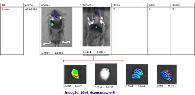
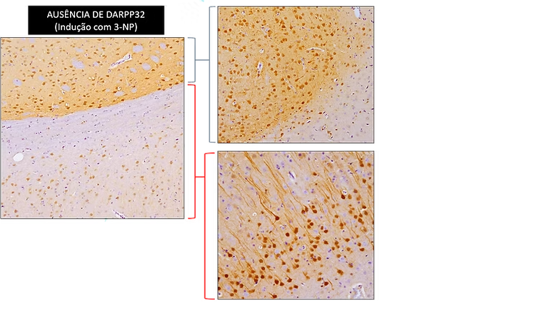
RESULTS OF CTIPD TRANSPLANT IN PRE-CLINICAL MODEL OF HUNTINGTON'S DISEASE INDUCED BY 3-NP (NITROPROPIONIC ACID)
CTIPD BIODISTRIBUTION IN 3-NP INDUCED HUNTINGTON'S DISEASE MODEL (AFTER 30 DAYS)
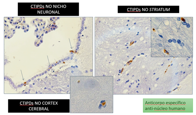
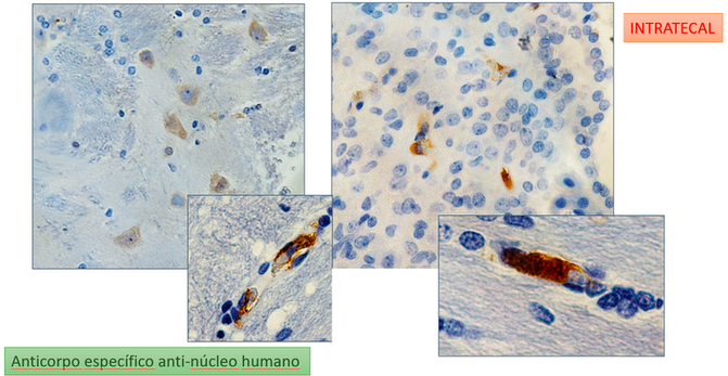
BIODISTRIBUTION STUDY CONDUCTED IN PARTNERSHIP WITH THE ALBERT EINSTEIN PRE-CLINICAL TESTING LABORATORY